Neurorehabilitation and Biomechanics Research Section

Pediatric Imaging and Neuroplasticity Team

Studies Actively Recruiting

Dopamine and Motor Learning in Cerebral Palsy
Protocol #16-CC-0149

The broad objective of this study is to determine the relationship between variations in genes related to dopamine (DA) neurotransmission in areas of the brain associated with motor leaning (e.g. DRD1, DRD2, DRD3, COMT, DAT) and/or to activity-dependent brain plasticity (e.g. BDNF) and differences in motor learning rates and cognitive processing abilities in persons with and without cerebral palsy (CP). We will also explore whether motor and cognitive learning abilities are correlated within individuals which could suggest similar underlying neural mechanisms. Finally we would like to evaluate the effect of rewards on procedural learning in those with and without CP, to preliminarily assess how behavioral manipulations of the DA system may affect learning.

This protocol will consist of an observational trial whereby subjects with and without CP will participate in two different training paradigms, one that involves learning novel working memory tasks and one that involves motor skill learning in the lower extremities. All will have blood draws for genetic analyses at baseline, the results of which will be related to changes in performance (learning) per task after training. A second part will be a within-subjects evaluation on the effects of reward (versus no-reward) during learning, which is presumed to increase dopamine transmission.

custom designed fNIRS probe
An image of our custom designed fNIRS probe utilized to measure activity from the motor areas of the brain during movement execution.
 

Functional Near-Infrared Spectroscopy (fNIRS) to Assess Brain Activity in Children and Adults with Movement Disorders
Protocol #13-CC-0110

Neural imaging during movement has become more portable by utilizing an approach termed functional near-infrared spectroscopy (aNIRS) as a means to isolate areas of brain activity. fNIRS is a non-invasive imaging technology that uses low levels of nonionizing red and near-infrared colored light to measure the hemodynamic state of the brain. Although use of these technologies for assessing cortical activation patterns is increasing, validation of these approaches, particularly in children and those with brain injuries such as cerebral palsy, is in the early stages. In this protocol, our objective is to systematically compare cortical activation patterns associated with specified motor and sensory tasks in healthy children and adults to those with unilateral or bilateral childhood-onset neurological injury. The results of this study are expected to increase knowledge of brain activation patterns across tasks and groups with and without brain injuries and to provide for future clinical studies with these technologies.

Typical Development and Cerebral Palsy
An example of a cortical activation map created from fNIRS. On the left is the activation of a typically developing child during left ankle dorsiflexion, showing the canonical contralateral response. On the right is the activation from a child with cerebral palsy, demonstrating more diffuse, and primarily ipsilateral, response.

fNIRS for Assessment of Early Peripheral Nerve Injury on Motor Cortex Development

The objective of this study is to investigate the effect of early peripheral nerve injury on motor cortex function in humans. Obstetric Brachial Plexus Palsy (OBPP) is an injury in which nerves controlling one arm and hand are damaged perinatally, with approximately 25-33% of children having persist residual deficits. Common impairments include muscle weakness, abnormal posture, bony deformation, shoulder and elbow contractures, dislocations, and limb shortening. Standard treatments for chronic OBPP include both conservative and surgical options, although there is no compelling evidence to support either approach. One potential reason for the poor outcomes in some children with OBPP is subsequent alterations to the cortical substrate resulting from the peripheral injury. These alterations may impact rehabilitation treatment efficacy. We are investigating this possibility by using functional near-infrared spectroscopy (fNIRS) to characterize brain activation during several upper limb tasks performed by in adolescents with OBPP and age matched typically developing children. We are also using brain MRI to study the underlying structural components that may contribute to differences in brain activation. The implications of this work include reframing how rehabilitation therapy is delivered to children with peripheral nerve injury.

Rigid Body Database
Protocol #90-CC-0168

The purpose of this study is to utilize motion capture technology to accurately and precisely quantifying the different ways people control limb and whole body movements. This information will be used to develop a database on normal movements and adaptive movements of people who have diseases that affect the way they move. The database will serve as a tool to improve diagnosis and treatment of patients with movement-related problems. Electrical activity in the muscles also may be measured, using small metal electrodes attached to the surface of the skin with an adhesive bandage.
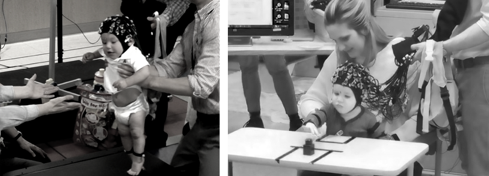
A 3D representation of a skeletal system.
An example of the biomechanical models constructed from our motion capture system which is used to precisely quantify movement.
 

Additional Areas of Investigation

  • Evaluation of an orthosis that uses electrical stimulation for foot drop in children with CP.
  • Using 3D motion capture and EMG to quantify and characterize different movement disorders such as dystonia in children and adults.
  • Detecting movement and balance problems in highly functional individuals who have sustained a brain injury using virtual reality technology.
  • Using ultrasound in patients and controls to study muscle architecture in both relaxed and contracted states.
  • Evaluating the kinematics and EMG patterns of different locomotor training devices compared to overground walking.

Completed Studies

Cerebral Palsy Coordination Protocol

This project started in Spring 2010 and evaluates lower extremity reciprocal coordination in ambulatory children with cerebral palsy (CP) across three levels of mobility and compared to children without CP. Children with CP are randomized to one of two interventions aimed to improve coordination: a motor-assisted cycle or an elliptical trainer. Both provide assistance and resistance to reciprocal leg movements. In the case of the elliptical, the assistance comes from the participant's own arm movements. Both interventions will be performed at a high cadence with resistance gradually introduced and progressed after target cadence is achieved. The implicit goal of this work is to determine whether intense reciprocal training of the lower extremities can stimulate sensorimotor pathways in CP to a sufficient degree to effect change in coordination and measurable changes in connectivity within and between the sensory and motor areas of the brain. The primary functional goal of the project is to help children with CP walk more quickly and efficiently by improving their ability to activate their leg muscles rapidly and reciprocally. This is the first clinical trial in CP using these devices in ambulatory children.

NOTE: PDF documents require the free Adobe Reader.

This page last updated on 01/17/2023

You are now leaving the NIH Clinical Center website.

This external link is provided for your convenience to offer additional information. The NIH Clinical Center is not responsible for the availability, content or accuracy of this external site.

The NIH Clinical Center does not endorse, authorize or guarantee the sponsors, information, products or services described or offered at this external site. You will be subject to the destination site’s privacy policy if you follow this link.

More information about the NIH Clinical Center Privacy and Disclaimer policy is available at https://www.cc.nih.gov/disclaimers.html