Radiology and Imaging Sciences

Colon carcinoma liver metastasis

Before ablation

CT scan of the liver before ablation shows large solitary lesion from colorectal carcinoma.

After treatment

Coronal reconstructed image after treatment shows dark treated area without contrast enhancement in the area of the tumor. The dark area is coagulative necrosis, or dead cells.

Reconstruction after ablation

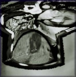
Three-dimensional reconstruction after ablation.

This colon cancer metastasis to the liver had been previously treated with surgery and cryotherapy but had recurred and failed chemotherapy.

NOTE: PDF documents require the free Adobe Reader.

This page last updated on 05/24/2022

You are now leaving the NIH Clinical Center website.

This external link is provided for your convenience to offer additional information. The NIH Clinical Center is not responsible for the availability, content or accuracy of this external site.

The NIH Clinical Center does not endorse, authorize or guarantee the sponsors, information, products or services described or offered at this external site. You will be subject to the destination site’s privacy policy if you follow this link.

More information about the NIH Clinical Center Privacy and Disclaimer policy is available at https://www.cc.nih.gov/disclaimers.html