
Content on this webpage is provided for historical information about the NIH Clinical Center. Content is not updated after the listed publication date and may include information about programs or activities that have since been discontinued.
The CC Imaging Sciences Program (ISP) will unveil the first phase of its new Picture Archiving and Communication System (PACS) and Radiology Information System (RIS) this month with the initiation of a sophisticated patient tracking system.
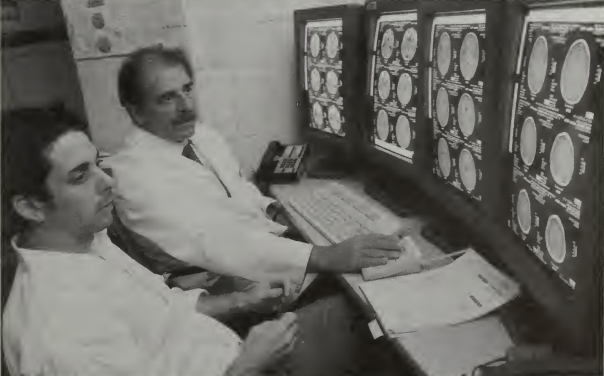
PACS is a high-speed, digital system for acquiring, storing, processing, and displaying images and reports. It will provide instant electronic access to current and past diagnostic images. RIS performs patient tracking and imaging-exam monitoring functions.
“This system will completely reengineer the operations of the radiology department,” said Jim Vucich, ISP manager and PACS/RIS project co-manager. “We expect it to increase the quality and efficiency of radiology services by reducing patient waiting times, improving image availability, and minimizing loss and misidentification of images and reports,” he said.
RIS is the first part of the system to be installed and will go on line for testing this month.
“For the first two or three months, we will still be maintaining data in paper form,” explained Vucich.
PACS is scheduled to begin operating within ISP in October and throughout NIH by next summer. RIS will track patient arrival and departure times, the start and end of exams, and when reports are dictated, read, and signed. Eventually, the system will include a voice recognition feature enabling spoken words to appear on the screen so reports can be immediately edited and verified. It also communicates with the medical information system (MIS) to receive orders for exams and send reports on completed exams back to the appropriate destinations.
Images stored in PACS/RIS originate from procedures and exams conducted in the Diagnostic Radiology, Nuclear Medicine, and PET Departments. They include CT scans, MR scans, PET scans, nuclear medicine scans, ultrasound examinations, and digital radiography examinations.
When an order for an imaging procedure is entered into MIS, the order will automatically be sent to RIS. The system will then track when the patient transport was called, when the transport left to transfer the patient, when the patient arrived in the radiology department, and when the patient entered and left locations within the department.
“Such detailed data will decrease patient waiting times and make better use of staff and equipment,” Vucich said.
Once an order for an imaging procedure is placed in the system, PACS/RIS will automatically retrieve prior scans and reports for that patient. The system integrates new and prior images and reports, so physicians can access and compare current images with previous scans and x-rays.
“This will make radiology images and reports more easily accessed by physicians, including after hours,” said Dr. Alberto Goldszal, chief, PACS/RIS section and PACS/RIS project co-manager.
It is customized to track images by protocol and institute. Radiologists will interpret images on large, high-resolution monitors in the Diagnostic Radiology Department. When the system is fully deployed next summer, patient images and their corresponding reports will be available on workstations and desktop computers across NIH through the use of the PACS web browser application. In addition, workstations dedicated to PACS/RIS image and report reviewing will be installed in specialized areas of the CC that conduct intensive image review activities. The images and reports will only be available to authorized users.
“This is the first step toward a filmless radiology department,” explained Vucich. “Eventually all images will be acquired, distributed, and stored in digital form. This will eliminate film and processing costs as well as lost and damaged films.”
“We expect this system to prove to be an efficient method for connecting people to the knowledge and resources they need at the appropriate times and locations for the best possible outcomes,” concluded Dr. Goldszal.
—by Colleen Henrichsen